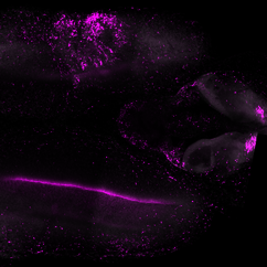
An atlas of the salamander brain and its regenerative abilities
An international team of scientists has mapped out all cell types in the salamander forebrain. In a colossal effort, they created an atlas of the axolotl forebrain and characterised the cells that give this animal the extraordinary ability to regenerate its neurons after an injury. Their results, alongside two other studies of the salamander brain, are now published in the journal Science.
When four-legged animals transitioned from an aquatic lifestyle to dwell on land millions of years ago, they were met with new challenges: how does one smell, learn, or memorise on land? The cognitive demands of a terrestrial lifestyle have led to evolutionary innovations in the brain. The layers of grey matter that cover the surface of the brain have grown more complex and have expanded into new regions. In mammals, this has resulted in the evolution of the neocortex, the large part of the brain that resembles a walnut’s tortuous surface. How the brain cells of ancestral land vertebrates have changed to give rise to the brains of reptiles, birds, and mammals is still a largely unanswered question.
One major piece of the brain evolution puzzle has been missing: amphibians. This diverse group of animals, comprising salamanders, frogs, and toads, without forgetting legless caecilians, branched off the vertebrate family tree about 350 million years ago, after the first vertebrates set fin on land, but long before mammals arose.
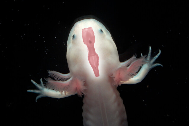
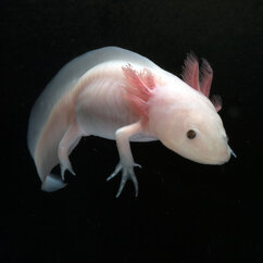
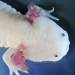
When it comes to investigating amphibian brains, the axolotl brings in some extra spice to the table: this Mexican salamander can fully regenerate parts of its brain after a major injury, a capacity that is inconceivable in terrestrial vertebrates. Mapping cell types in the axolotl brain and comparing them to those of other vertebrates could shed light on the origins of modern vertebrate brain regions and on the mechanisms underlying brain regeneration.
In a collaborative tour de force, scientists in the labs of Elly Tanaka at the IMP and Barbara Treutlein at ETH Zurich have created an atlas of the axolotl’s forebrain and its cell types. They have now published their research in the journal Science, alongside two other studies of the salamander brain.
Gallery
The salamander brain and relatives
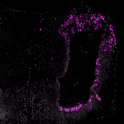
The researchers used single-nucleus sequencing, a popular genomics method to categorise cell types based on the genes they activate. They catalogued the types of neurons, stem cells, and progenitor cells that make up the axolotl forebrain. In parallel, another team led by Maria Tosches at Columbia University created a similar atlas for a different salamander species.
“In the last few years, scientists have analysed and compared the brain cell types of many animals with a spinal cord, from turtles to lizards, birds, mice, and primates. Nobody had looked at amphibians with this approach until now,” explains Katharina Lust, postdoc in the lab of Elly Tanaka and co-first author of the study alongside Ashley Maynard and Tomás Gomes from the lab of Barbara Treutlein. “Our work shows how the brain cell types in axolotls relate to known cells and regions in other animals.”
Lust and colleagues identified groups of neurons in the axolotl that match the mammalian hippocampus – a region of the brain involved in memory formation – and parts of the cortex involved in olfaction. Intriguingly one small corner of the brain showed possible similarity to the cerebral cortex. In future experiments, the team will aim to figure out whether the different brain regions in axolotl have similar connectivity and function to their counterparts in mammals. The axolotl will also represent an ideal model to study how these regions, after being injured, can recover their function through regeneration.
“Expanding our repertoire of brain atlases to older animal lineages, such as amphibians, fish, and lamprey, will allow us to understand which circuits were preserved through evolution and which ones changed as animals diversified,” says Elly Tanaka. “Charting cell types in the axolotl brain does not only bring evolutionary insights into the vertebrate brain, but also paves the way for innovative brain regeneration research.
Regenerating lost neurons
One type of cell is responsible for neuron production in the salamander central nervous system: ependymoglia cells. Throughout an axolotl’s life, ependymoglia cells divide and differentiate to generate new neurons. They also take care of regenerating lost neurons after an injury.
In their study, the team categorised ependymoglia cells and characterised the state they enter to launch regeneration. Should the brain suffer an injury, some of these cells activate an injury-specific set of genes and eventually regenerate lost neurons.
These results match those of another, independent study on the mechanisms of axolotl brain regeneration, published in the same Science issue. This research was led by former IMP postdoc Ji-Feng Fei, now a group leader at the Guangdong Academy of Medical Sciences, China.
“Our ultimate goal is to understand what brain stem cells do after an injury – what genes they activate, how they interact, and how they eventually recreate neurons that will recover the lost connections. How does each cell ‘know’ what do to? We will keep working on these questions with our collaborators in Switzerland,” says Lust.
“This paper highlights the power of collaboration,” says Barbara Treutlein, Professor at the ETH Zurich. “Elly Tanaka and her lab at the IMP brought in their expertise in experimental biology with the axolotl, and we added our extensive experience in single-cell genomics and computational biology to publish our findings in record time. We look forward to continuing this fruitful collaboration to reveal the secrets of brain regeneration.”
Original publication
*These authors contributed equally to this study.
Further reading
The axolotl, new model for bone fracture healing
